
Caring for the Health of Your Eyes
New Technology Leads to Earlier Diagnosis.
The new technology at Lifetime Eye Care allows us to detect Macular Degeneration, Glaucoma, Diabetic Retinopathy, and other abnormalities sooner than ever before, thus making early intervention possible.
Oculus Keratograph 5M
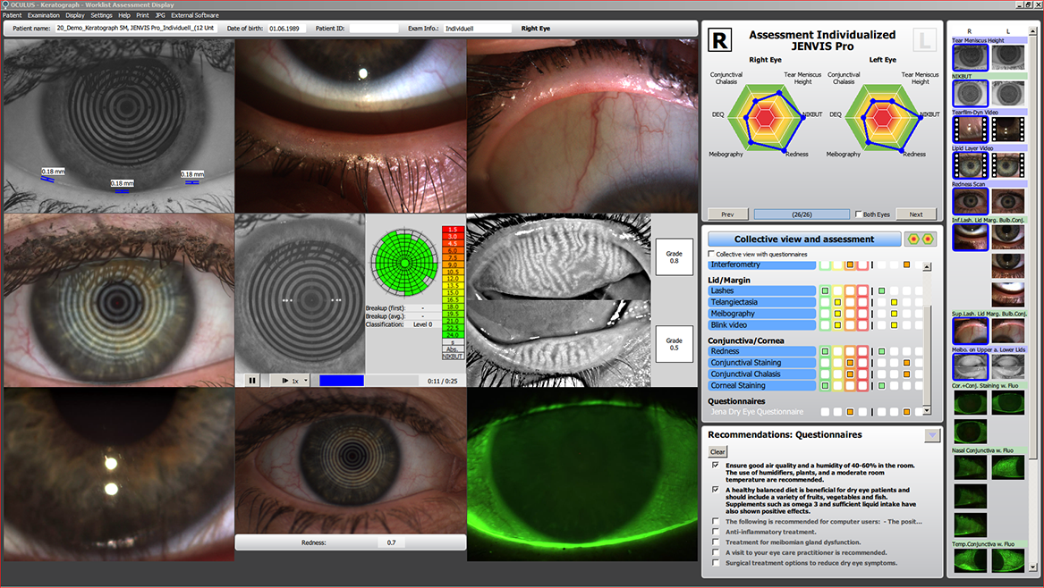
Our new dry eye equipment allows for a more thorough investigation into the cause of each patient’s unique dry eye disease, allowing our doctors to select the best treatments available for each scenario. Meibography, or non-invasive imaging of the oil glands within the eyelids (called the Meibomian glands), is one of the features of this new technology. Meibomian Gland Dysfunction is a leading cause of dry eye amount in the adult population and current studies show this condition is starting younger in the age of digital devices. Schedule your dry eye evaluation with one of our doctors today!
Specular Microscopy (Nidek CEM-530)

Ever wonder why contact lenses are considered medical devices or why eye doctors request to see contact lens wearers at least every year? The truth is, placing a contact lens on the surface of the eye does not always come without consequences. One such complication involves changes to the eye itself at a cellular level. Specular microscopy allows our doctors to look for microscopic changes in the cornea (the clear windshield on the front of the eye) that can be induced by contact lens wear. Some of these changes, if left unchecked, can result in permanent vision loss. This simple, non-invasive scan is run on every contact lens wearer that walks through our doors each year to monitor for changes in eye health related to contact lens wear, and it takes less than one minute!
Dark Adaptation
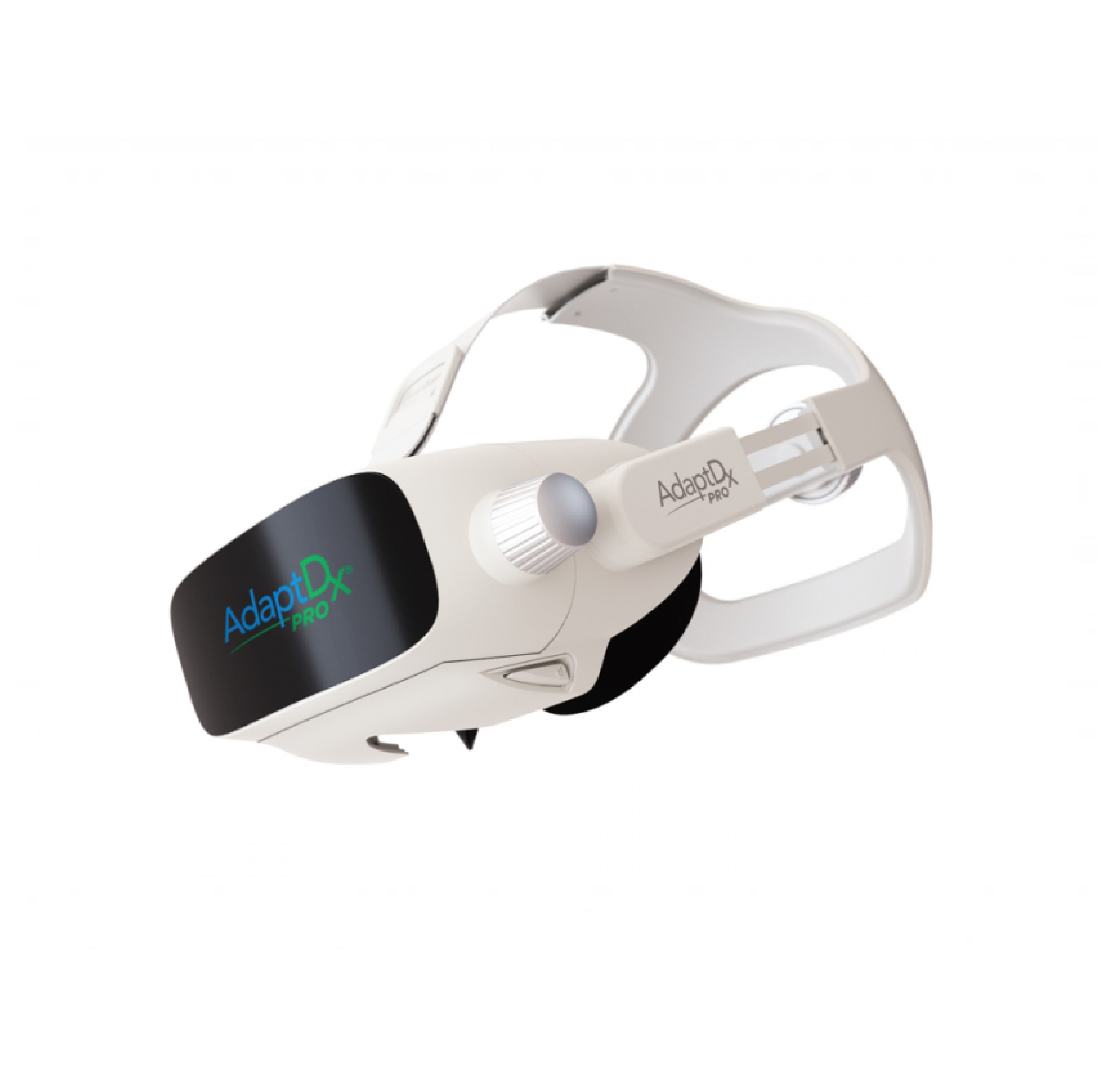
The Adapt Dx (Maculogix) is based on the functional test of dark adaptation—that is, the transition from light situations to darkness. Research has proven that the darkness adaptation of our vision is impaired up to 4 years before Macular Degeneration is clinically relevant.
The screening test is non-invasive and can be completed within 5 minutes or less by a technician. If results are found to be abnormal, a more extensive threshold test may be performed.
Fundus Imaging with Autofluorescence
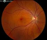 Fundus autofluorescence (FAF) is a non-invasive technique to help analyze the health of the retina. Hence, it serves as a precursor to retinal damage associated with diabetes, macular degeneration and other diseases.
Any routine eye exam includes a careful look at the retina, which is located at the back of the eye, to screen for abnormalities or disease. The sensitive tissue of the retina is susceptible to a variety of diseases that can ultimately lead to partial loss of vision or complete blindness. Early detection of any retinal abnormality is crucial.
Optomap Retinal Imaging is utilized diagnostically at Lifetime Eye Care for all patients as we attempt early detection which may mean early intervention. It may also mean starting patients sooner on vitamin therapy in hopes of preventing the decline to advanced Macular Degeneration. It may also mean more frequent monitoring for patients found to have Diabetic Retinopathy or Macular Degeneration by these new diagnostic measures. Ultimately, these new techniques and equipment will hopefully help our patients maintain better vision and a better quality of life as they age.
OCT Imaging
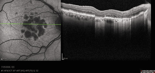 Optical coherence tomography (OCT) is a non-invasive imaging test that uses light waves to take cross-section pictures of your retina, the light-sensitive tissue lining the back of the eye.
With OCT, each of the retina’s distinctive layers can be seen, allowing Drs. Duzan, Hennig, and Esarey to map and measure their thickness. These measurements help with diagnosis and provide treatment guidance for glaucoma and retinal diseases, such as age-related macular degeneration and diabetic eye disease.